In this post Dr Thomas Woolley reviews a recent conference he organised in Oxford. Bringing together experimentalists and theoreticians from around the world they had one focus: blebbing cells!
If you have never heard of blebbing, you can be forgiven. Indeed, for a long time blebs were completely ignored as it was thought that they were signs of cell death. However, over the last twenty years experimentalists and theoreticians have been working hard to change the image of the humble bleb, from a simple nuisance to a sign of incredibly complex dynamics. This conference brought together the world’s leading experts in blebbing with the hope of not just simply offering new insights but also the chance to develop new contacts and breed new interdisciplinary collaborations.
To understand what a bleb is we must first understand a little about the structure of a cell. At its simplest, the cell can be characterised as an extremely thin and weak membrane which is attached to a stiffer actin cortex through adhesion proteins. These structures surround a pressurised fluid, known as cytosol. If the membrane becomes unstuck from the cortex, the intercellular pressure is able to push the membrane into a hemispherical blister, known as a bleb. Over time, cortex reforms in the bleb, which is then slowly retracted (see Fig 1). Although this may sound simple, certain cells are able to produces these protrusions rapidly and randomly all over the membrane (see Fig 2) and the processes that control each of these stages is not fully understood.
 |
| Figure 2. A single cell that has produced a number of blebs. Electron microscopy photo courtesy of the Skeletal Muscle Development Group, University of Reading. |
Now-a-days blebs are known to aid in cell spreading, division, motility and many other processes besides. It was with this knowledge that experts from each of these blebbing fields were gathered together in the brand new Mathematical Institute of the University of Oxford. If you are wondering why we were in the mathematical department and not the biology department, it is simply because blebbing has a proud tradition of being a very multidisciplinary research area. Many of the field’s leading papers have both theoretical and experimental authors demonstrating what can be achieved when biological insights are tested mathematically, which, in turn leads to new predictions for experimentation.
The conference was opened with a wonderful plenary talk delivered Ewa Paluch. Being one of the field’s leading experimentalists she masterfully wove together the complex history of blebbing with modern insights, finally leading to new exciting work dealing with cells that are able to rapidly oscillate between bulging states. Her talk was then followed by Guillaume Salbreaux, one of her theoretical collaborators, who presented a number of mathematical frameworks in which these experiments could be understood.
Different blebbing cell were introduced by Ketan Patel and Henry Collins-Hooper, who demonstrated that the healing of muscle damage depends crucially on the ability of muscle stem cells to bleb. Working with Thomas Woolley of the Oxford Mathematical Institute, who also spoke, they were able to produce some ground breaking conclusions on how to regenerate the blebbing characteristics of older cells, thereby potentially allowing wounds to heal quicker. Phil Dash then presented the next stages of their work, involving a new direction considering membrane pores called “aquaporins”. Aquaporins let water in and out of the cell and have the potential to revolutionise the way we control bleb location and therefore cell motion.
One of the most exciting presentations of the day was from Robert Grosse. Robert surprised everyone by showing recent experiments of cells that were able to use blebs to invade other cells. His results were so new that there were gasps of amazement from the audience!
Another theoretical/experimental team from Cancer Research UK then presented some beautifully realistic simulations of blebbing cells and there corresponding experimental analogues. The work of Paul Bates, Melda Tozluoğlu and Erik Sahai, recently published in Nature Cell Biology, has set a new precedent, by which all new work will be measured. In particular they characterised why different methods of motility help the cell travel faster in different environments.
The second day was also predominantly about blebbing, with talks from Guillaume Charras on the adhesions that fix the membrane and cortex together and Wanda Strychalski presenting a simple but effective mathematical model that tests different assumptions about the fluid interior. The day also saw some more general cellular motion and membrane dynamic talks from Laura Kimpton and Marrino Arroyo, respectively. Laura’s prize winning work considers the crawling dynamics of cells and has much potential to be extended. Marrino’s cutting edge numerical methods offered simple reasons behind extremely complex membrane protrusions.
 |
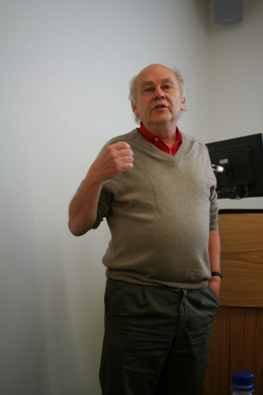
| Figure 4. Robert Kay, MRC Laboratory of Molecular Biology, speaking with passion about blebs in dictyostelium cells. |
The final talk by Robert Kay once again challenged our preconceived notions of cellular motion. He supplied convincing evidence suggesting a link between blebs and other cellular protrusions known as “lamellipodia”. Previously, it had been assumed that lamellipodia were quite different to blebs as lamellipodia were pushed out by the actin cortex, whilst blebs are simply driven by the pressure difference. However, Robert’s work suggests that the membrane of the lamellipodia may also be pushed out due to pressure gradients, whilst the actin skeleton fills in the space behind the membrane, thereby not actually pushing the membrane at all!
The two days were a wonderful meeting of minds. The focused direction of all of the participants led to much discussion of new ideas, new twists of old ideas and hopefully new collaborations. For myself, the take home message from the conference was that although we may all refer to our membrane protrusions as blebs, the protrusions come in many different shapes and sizes. Some look like small spherical hemispheres that grow randomly over the entire cell, whilst others are large amorphous expansions that are able to flow along chemical gradients.
Whatever the future may hold for blebs and membrane dynamics, it is certain that we are still generating more questions than answers.


Can you tell us more about the talk from Robert Grosse? What cells invade what cells, and what for?
ReplyDeleteI was diagnosed as HEPATITIS B carrier in 2013 with fibrosis of the
ReplyDeleteliver already present. I started on antiviral medications which
reduced the viral load initially. After a couple of years the virus
became resistant. I started on HEPATITIS B Herbal treatment from
ULTIMATE LIFE CLINIC (www.ultimatelifeclinic.com) in March, 2020. Their
treatment totally reversed the virus. I did another blood test after
the 6 months long treatment and tested negative to the virus. Amazing
treatment! This treatment is a breakthrough for all HBV carriers.